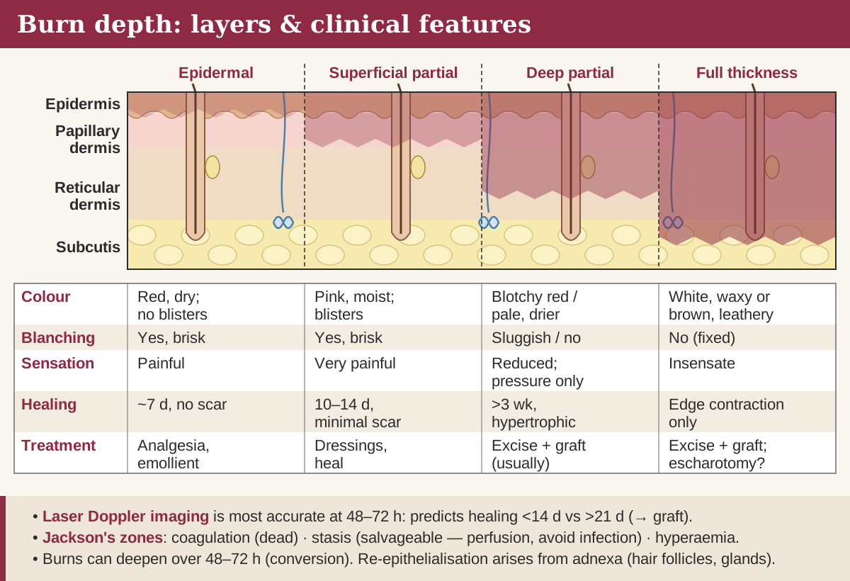
Burn depth by skin layer, with the clinical features of each.
Burn depth: what you see and what it predicts
Depth is classified by how much dermis survives, and its practical job is to predict whether the wound will heal within about three weeks. Burns that take longer carry a high risk of hypertrophic scarring and are usually excised and grafted.
Clinical features of burn depth. Most burns are mixed; reassess at 48–72 hours.
Depth
Appearance
Capillary refill
Sensation
Healing
Epidermal (superficial)
Red, dry, no blisters
Brisk
Painful
About 7 days, no scar
Superficial partial thickness
Pink, moist, blistered
Brisk
Very painful
10–14 days, minimal scarring
Deep partial thickness
Blotchy red or pale, drier; fixed staining
Sluggish or absent
Reduced; pressure only
Over 3 weeks; usually excise and graft
Full thickness
White and waxy, or brown and leathery
None
Insensate
Only by contraction from the edges; excise and graft
Depth is dynamic, and Jackson's zones explain why. A central zone of coagulation is already dead; a surrounding zone of stasis is under-perfused but salvageable; an outer zone of hyperaemia will recover. Hypovolaemia, infection, oedema and cooling tip stasis into necrosis, so a burn can deepen over the first 48–72 hours. Partial-thickness burns heal from surviving adnexa — hair follicles and sweat glands — which is why deep dermal burns heal slowly. Laser Doppler imaging after about 48 hours predicts healing potential better than clinical judgement alone.
Estimating TBSA
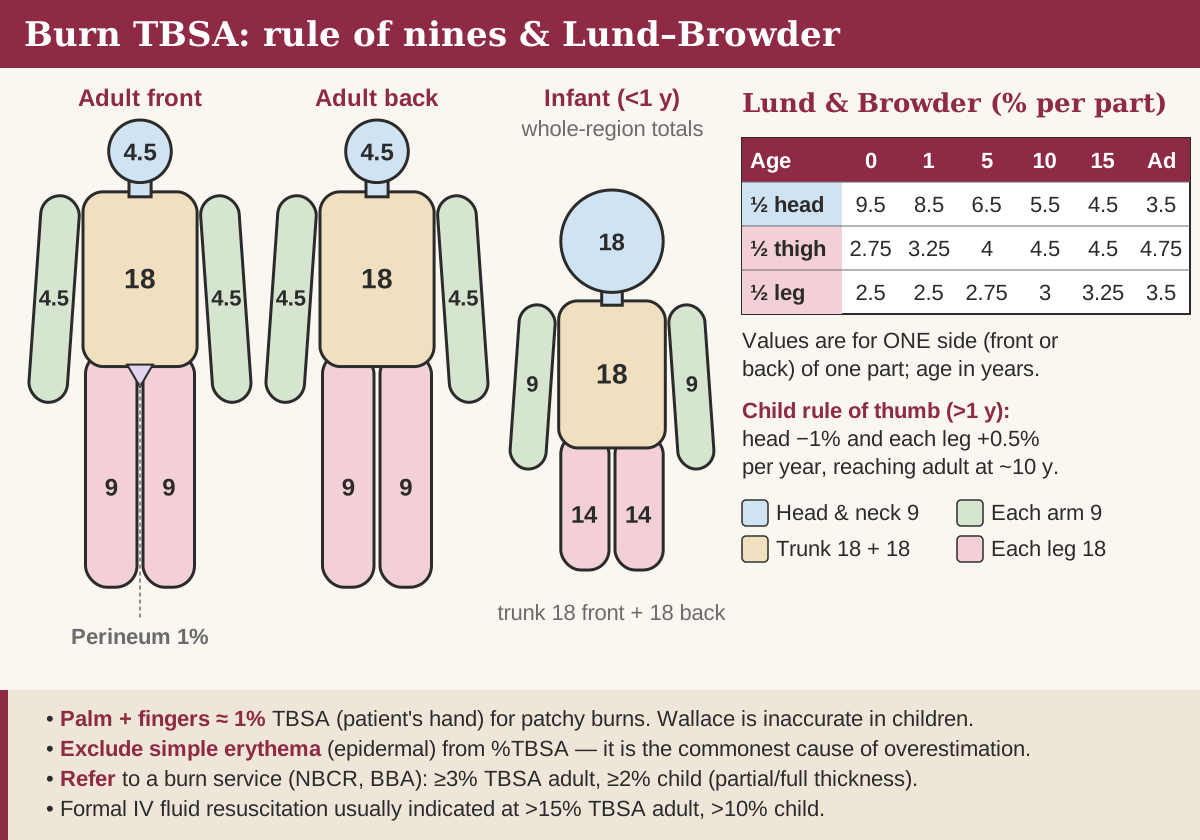
Rule of nines for adults and infants, with Lund–Browder age adjustments.
Only partial- and full-thickness burns count towards TBSA. Simple erythema is excluded, and including it is a classic cause of overestimating small burns and starting unnecessary resuscitation.
Wallace rule of nines (adults): head and neck 9%, each arm 9%, front of trunk 18%, back of trunk 18%, each leg 18%, perineum 1%.
Palmar method: the patient's palm with adducted fingers is about 1% TBSA — useful for small or scattered burns, and for very large burns by estimating the unburnt skin.
Lund–Browder chart: the most accurate method and the standard in children, because it adjusts head and leg proportions for age.
In infants the head is about 18% and each leg about 14%. For each year after the first, take roughly 1% from the head and add 0.5% to each leg, reaching adult proportions at around ten years.
Lund–Browder age-variable areas: % TBSA for one surface (front or back) of each part.
Age (years)
½ of head
½ of one thigh
½ of one lower leg
0
9.5
2.75
2.5
1
8.5
3.25
2.5
5
6.5
4
2.75
10
5.5
4.5
3
15
4.5
4.5
3.25
Adult
3.5
4.75
3.5
Who to refer to a burns service
The National Burn Care Referral Guidance used in England and Wales sets deliberately low thresholds, judged on size, depth, site, mechanism and patient. Refer:
Burns of 3% TBSA or more in adults, or 2% or more in children
All full-thickness and all circumferential burns
Any burn not healed within 2 weeks
Any burn where non-accidental injury is suspected
Discuss with a burns consultant: burns of the face, hands, feet, perineum or genitalia; chemical, electrical, friction and cold injuries; an unwell or febrile child with a burn; and comorbidities that may affect treatment or healing. Suspected inhalation injury is discussed too, once the airway is safe. Older UK and international lists use higher size thresholds — 10% TBSA in adults, 5% in children, full thickness over 5% — and still appear in some texts; when a question names the current national guidance, answer with it.
Fluid resuscitation
Formal intravenous resuscitation is usually started for burns over 15% TBSA in adults and over 10% in children. The Parkland formula estimates the first 24 hours of crystalloid — classically 4 mL × weight (kg) × %TBSA of Hartmann's solution — with half in the first 8 hours from the time of injury and the rest over the following 16.
8,400 mL
24-hour Parkland estimate: 4 × 70 kg × 30% TBSA
≈525 mL/h
first 8 hours from injury (4,200 mL)
≈263 mL/h
next 16 hours (4,200 mL)
The formula is a starting estimate, not a prescription. Patients often end up receiving more than it predicts, so many UK units start lower — 2–3 mL/kg/%TBSA, as in the modified Brooke formula — and titrate hourly against urine output. Children also need glucose-containing maintenance fluid on top of resuscitation, calculated by Holliday–Segar (100 mL/kg/day for the first 10 kg, 50 mL/kg/day for the next 10 kg, 20 mL/kg/day thereafter), with blood glucose checked.
Endpoints and fluid creep
Urine output is the main endpoint: aim for 0.5–1 mL/kg/h in adults and about 1 mL/kg/h in children, measured hourly through a urinary catheter. Low output with tachycardia, rising haematocrit and lactate means under-resuscitation — increase the crystalloid rate rather than reaching for a diuretic or vasopressor in a hypovolaemic patient.
Over-resuscitation is the opposite danger. Fluid creep causes pulmonary oedema, abdominal and limb compartment syndromes and deepening of the burn from oedema, so output persistently above target means the rate should come down. Electrical injury with myoglobinuria is the exception that needs a higher target, around 1–2 mL/kg/h in adults, until the urine clears.
Inhalation injury, carbon monoxide and cyanide
Suspect inhalation injury after any fire in an enclosed space and secure the airway early, because oedema worsens over the first 24–48 hours and can make later intubation impossible. Stridor is a late sign; do not wait for it.
Facial burns, singed nasal hair or eyebrows, soot in the mouth or nose, carbonaceous sputum
Confusion or reduced consciousness, which may reflect carbon monoxide or cyanide
Carbon monoxide
Binds haemoglobin far more avidly than oxygen and shifts the dissociation curve to the left. Pulse oximetry reads falsely normal, so measure carboxyhaemoglobin by co-oximetry. Give 100% oxygen at once: it cuts the half-life from about 4 hours on air to roughly 60–90 minutes.
Hydrogen cyanide
Released from burning plastics, foam, wool and silk. It blocks cytochrome c oxidase, causing histotoxic hypoxia with a high lactate despite adequate oxygen delivery. Treat with intravenous hydroxocobalamin, which binds cyanide as cyanocobalamin for renal excretion; it turns skin and urine red and interferes with some laboratory assays.
Escharotomy
Escharotomy is indicated when circumferential deep dermal or full-thickness eschar threatens perfusion of a limb or restricts ventilation. Inelastic eschar cannot expand as oedema builds beneath it, so it acts like a tourniquet.
Limbs: mid-medial and mid-lateral incisions through the eschar into fat until it springs apart — not through deep fascia. Protect the ulnar nerve behind the medial epicondyle and the common peroneal nerve at the fibular neck.
Chest: incisions along both anterior axillary lines joined by a transverse cut across the costal margins, releasing a ‘breastplate’ when airway pressures rise and tidal volumes fall.
After release: if perfusion does not return, or the injury is electrical or crushing, suspect a true subfascial compartment syndrome and perform fasciotomy.
A 70 kg man is 4 hours into resuscitation for a 40% TBSA flame burn. His urine output over the last hour was 12 mL, he is tachycardic at 120/min, his haematocrit is rising and his serum lactate is climbing; his blood pressure is maintained. The prescribed resuscitation and maintenance fluids are being delivered exactly as calculated.
What is the most appropriate management?
How sure are you?
Explanation
The picture is under-resuscitation: urine output well below the 0.5-1.0 mL/kg/hr target (12 mL/hr is about 0.17 mL/kg/hr) with tachycardia, haemoconcentration and a rising lactate. Burns formulae only estimate the starting rate, so fluids must be titrated up to the urine-output endpoint, and the correct step is to increase the crystalloid resuscitation rate. A vasopressor treats vasodilatation, not the hypovolaemia of burn oedema, and can worsen perfusion; furosemide is dangerous when the patient is under-filled; making no change ignores that the formula is only a guide; and switching to 0.9% saline risks hyperchloraemic acidosis without addressing volume. Reference: EMSB; British Burn Association resuscitation endpoints.
A 34-year-old man is rescued from a house fire in an enclosed room. He has burns to the face, singed nasal hair and carbonaceous sputum, and his voice has become hoarse; there is progressive swelling of the lips and neck. He maintains oxygen saturations of 99% on high-flow oxygen and has no stridor.
What is the most appropriate management?
How sure are you?
Explanation
This describes an evolving upper-airway inhalation injury with impending obstruction: facial burns, singed vibrissae, carbonaceous sputum, hoarseness and progressive supraglottic oedema. EMSB/ATLS teaching is to secure the airway early with elective endotracheal intubation before oedema (which worsens over the first 24-48 hours) makes intubation impossible; hoarseness signals laryngeal involvement and a normal saturation is falsely reassuring. Waiting for stridor (D) is dangerous because stridor is a late pre-obstructive sign; nebulised adrenaline and steroids (A) and observation (B) do not arrest progressive oedema; deferring an awake technique until fasted (E) wastes the safe window in an emergency. Reference: EMSB airway guidance.
Multiply 4 mL by weight in kg by %TBSA to get the 24-hour volume of Hartmann's. Give half in the first 8 hours from the time of injury and half over the next 16, then titrate to urine output.
Should erythema be included in TBSA?+
No. Only partial- and full-thickness burns count; including erythema inflates the estimate and can trigger unnecessary intravenous resuscitation.
What urine output should a burns resuscitation aim for?+
About 0.5–1 mL/kg/h in adults and around 1 mL/kg/h in children. Output persistently above target means the rate should be reduced to avoid fluid creep.
When is escharotomy needed?+
When circumferential deep or full-thickness eschar compromises limb perfusion or chest wall movement. It releases the eschar, not the fascia; fasciotomy is reserved for true compartment syndrome.
Why is pulse oximetry misleading in carbon monoxide poisoning?+
Standard oximeters cannot distinguish carboxyhaemoglobin from oxyhaemoglobin, so saturations look normal. Carboxyhaemoglobin must be measured by co-oximetry.
References
British Burn Association. Emergency Management of Severe Burns (EMSB) course manual.
National Network for Burn Care. National Burn Care Referral Guidance, 2012.
Janis JE (ed). Essentials of Plastic Surgery, 3rd ed. Thieme — chapters on burn assessment and acute burn management.
Richards AM, Dafydd H. Key Notes on Plastic Surgery, 2nd ed. Wiley-Blackwell — Burns.
Chung KC (ed). Grabb and Smith's Plastic Surgery, 8th ed. Wolters Kluwer — chapters on thermal injury.
Written for revision in the FRCS (Plast) style under our editorial standards. It is not clinical advice: always confirm against current guidance. Spotted an error? Tell us.