Flexor tendon injuries: zones, pulleys, repair and rehabilitation
Flexor tendons for FRCS (Plast): Verdan zones, A2/A4 pulleys, Camper's chiasm, examination, four-strand repair, early active motion, rupture and Leddy–Packer.
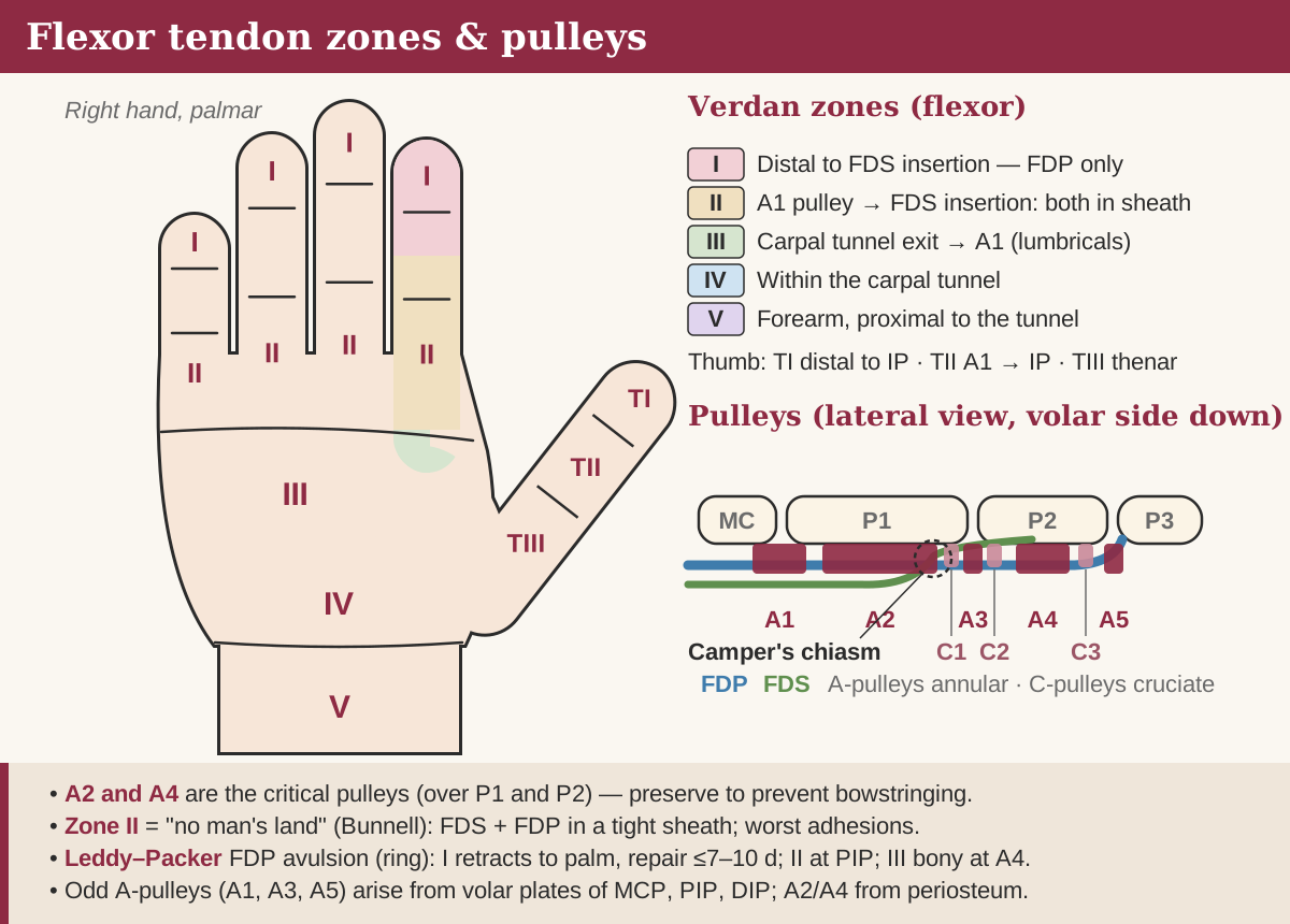
Verdan flexor zones, the finger pulley system and Camper's chiasm.
Verdan's zones
Flexor injuries are described by Verdan's five zones, from fingertip to forearm, and the zone predicts both the anatomy you will find and the outcome you can expect.
Verdan flexor zones of the hand and thumb.
Zone
Boundaries
What lies there
I
Distal to the FDS insertion
FDP alone; includes avulsion (jersey finger)
II
Proximal edge of the A1 pulley to the FDS insertion
FDS and FDP together in the fibro-osseous sheath — ‘no man's land’
III
Distal edge of the carpal tunnel to the A1 pulley
Lumbricals arising from FDP; superficial palmar arch; common digital nerves
IV
Within the carpal tunnel
Nine flexor tendons and the median nerve beneath the transverse carpal ligament
V
Proximal to the carpal tunnel
Musculotendinous junctions; median and ulnar nerves; radial and ulnar arteries
T1–T3 (thumb)
T1 distal to the IP joint; T2 A1 pulley to IP joint; T3 thenar eminence
FPL in its sheath (T1–T2); FPL among the thenar muscles (T3)
Zone II earned the name ‘no man's land’ because two tendons glide against each other inside a tight sheath, so adhesions between them and to the sheath are hard to prevent. Tang subdivides it (IIA–IID) to guide decisions about pulley venting and FDS repair.
FDS, FDP and Camper's chiasm
FDS flexes the PIP joint and FDP flexes the DIP joint, and their relationship changes along the finger. Over the proximal phalanx FDS splits into two slips that pass around FDP and reunite dorsal to it at Camper's chiasm before inserting on the middle phalanx; FDP passes through the split to insert at the base of the distal phalanx.
Innervation: FDS is supplied by the median nerve; FDP to the index and middle fingers by the anterior interosseous nerve and to the ring and little fingers by the ulnar nerve; FPL by the anterior interosseous nerve.
Shared belly: FDP to the middle, ring and little fingers shares a common muscle belly while index FDP is largely independent — the basis of the quadriga effect when one FDP is tethered or over-advanced.
Nutrition: inside the sheath tendons are fed by synovial diffusion as well as by the vincula longa and brevia; diffusion matters most in zone II.
Healing: intrinsic healing by tenocytes competes with extrinsic healing from the sheath, which brings adhesions; early motion favours the intrinsic route.
The pulley system
Each finger has five annular (A1–A5) and three cruciate (C1–C3) pulleys, and A2 and A4 matter most: they hold the tendons against bone and prevent bowstringing. The odd-numbered annular pulleys (A1, A3, A5) arise from the volar plates of the MCP, PIP and DIP joints; A2 and A4 arise from the periosteum of the proximal and middle phalanges. The thin cruciate pulleys lie between them and collapse to allow flexion.
The thumb has its own arrangement — A1 over the MCP joint, an oblique pulley over the proximal phalanx and A2 near the IP joint — and the oblique pulley is the critical one. In the fingers, limited venting of A4 or part of A2 is now accepted when a bulky repair would otherwise catch, provided the rest of the system is intact.
Interactive 3D · Flexor tendon sheath & pulleys. Find A2 over the proximal phalanx and A4 over the middle phalanx, and see why losing them lets the tendons bowstring.Open full screen
Examination
Start with posture: a finger lying out of the normal flexion cascade at rest suggests a divided tendon before you touch it. Then test each tendon in isolation and always check the digital nerves, which run beside the sheath and are often injured with zone II lacerations.
FDP: hold the PIP joint extended and ask for DIP flexion.
FDS: hold the other fingers fully extended to neutralise the shared FDP belly, then ask the patient to flex the finger; PIP flexion confirms an intact FDS.
Tenodesis: passive wrist extension normally flexes the fingers; a finger that stays straight has lost its flexors.
Partial division: pain on resisted flexion or triggering — explore any suspicious wound.
Repair principles
Repair as a primary or early delayed primary procedure with atraumatic handling, a core suture of at least four strands and a running epitendinous suture. Strength rises roughly in proportion to the number of strands crossing the repair, and a four- or six-strand core is strong enough to tolerate early active motion. Late repair becomes progressively harder as the tendon retracts, softens and the muscle shortens.
Exposure: Bruner zigzag or mid-lateral incisions that incorporate the wound; retrieve a retracted proximal end gently, for example by catheter delivery, rather than by repeated blind grasping.
Core purchase of roughly 7–10 mm on each side, tensioned so the repair bunches by about 10%. This pre-tension resists gapping; gaps of more than about 3 mm weaken the repair and invite adhesions.
Epitendinous suture: a fine running monofilament adds strength and smooths the repair so it glides beneath the pulleys.
Both tendons in zone II are usually repaired; excising one slip of FDS can relieve crowding distally.
Partial lacerations of less than about half the cross-section can be trimmed rather than sutured.
Zone I: with a short distal stump, reattach FDP to bone; avoid advancing it more than about 1 cm, which risks quadriga and a flexion contracture.
Rehabilitation, rupture and outcomes
Early active motion is the standard after a strong four-strand repair: supervised by a hand therapist, it limits adhesions without a meaningful rise in rupture risk. The hand is protected in a dorsal blocking splint — wrist around neutral to slight flexion, MCP joints flexed, IP joints straight — and controlled active flexion starts within the first few days.
A typical progression after zone II repair with early active motion; local protocols vary.
Time after repair
Typical stage
0–6 weeks
Splint; protected active and passive flexion; no gripping
About 6 weeks
Splint discarded; light functional use
About 8 weeks
Graded resisted exercise
About 12 weeks
Unrestricted heavy loading and forceful grip
Passive regimes — Kleinert (active extension against rubber-band flexion) and Duran (controlled passive motion) — remain options for patients who cannot follow an active programme. Reported rupture rates after modern repair and early motion are in single figures, commonly quoted at around 4–10%; most ruptures occur during the early softening phase or with unguarded use and should be re-repaired promptly. Zone II has the least predictable range of motion. Adhesions that limit active but not passive flexion are treated by tenolysis, usually no earlier than about three months, once the soft tissues are supple and passive motion is maximised.
Jersey finger: Leddy–Packer
A jersey finger is an avulsion of FDP from the distal phalanx when an actively flexing finger is forced into extension — classically grabbing an opponent's shirt — and the ring finger is the usual victim. The patient cannot flex the DIP joint. Leddy and Packer classify it by how far the tendon retracts, which determines the vincular supply and the urgency of repair.
Leddy–Packer classification of FDP avulsion.
Type
Findings
Management
I
Tendon retracts into the palm; both vincula ruptured
Urgent reinsertion, ideally within 7–10 days, before the muscle shortens and the tendon degenerates
II
Tendon retracts to the PIP joint, held by the vinculum longum; may carry a small bony fleck
Reinsertion; the preserved vincular supply makes later repair more feasible, but earlier is better
III
Large bony fragment caught at the A4 pulley; the tendon cannot retract further
Open reduction and fixation of the fragment
IV
Bony fragment, with the tendon also avulsed from it and retracted
A 32-year-old scaffolder is six weeks after four-strand zone II repair of both flexor tendons of the index finger, rehabilitated with an early active regime. The splint has been discarded and he has good active flexion without triggering. He asks when he can resume heavy lifting and forceful gripping.
At what point after repair is unrestricted heavy loading most appropriate?
How sure are you?
Explanation
This is return to work after flexor tendon repair. The splint is typically discarded at around six weeks, with light functional use from then, graded resisted exercise from about eight weeks, and unrestricted heavy or forceful gripping deferred until about twelve weeks, when the repair has regained enough tensile strength to make late rupture unlikely. Returning now or at eight weeks exposes a still-remodelling repair to peak loads; and delaying to sixteen weeks or six months is unnecessary for an uncomplicated repair and prolongs time off work. Reference: Janis, Essentials of Plastic Surgery 3e, ch. 80 (postoperative care and rehabilitation).
The segment from the proximal edge of the A1 pulley to the FDS insertion, where FDS and FDP run together inside the fibro-osseous sheath. Adhesions are common there, so outcomes are the least predictable.
Which flexor pulleys are most important?+
A2 and A4 in the fingers, because they hold the tendons against bone and prevent bowstringing. In the thumb, the oblique pulley.
How many strands should a flexor tendon repair have?+
At least four core strands, combined with an epitendinous suture, to withstand early active motion.
When can a patient return to heavy lifting after flexor tendon repair?+
Usually at about 12 weeks. The splint typically comes off at around 6 weeks and graded resisted exercise starts at around 8.
What is the Leddy–Packer classification?+
It grades FDP avulsions by retraction: type I into the palm, type II to the PIP joint, type III with a large fragment held at A4, and type IV with a fragment plus a tendon that has pulled off it and retracted.
References
Janis JE (ed). Essentials of Plastic Surgery, 3rd ed. Thieme — chapter on flexor tendon injuries and rehabilitation.
Richards AM, Dafydd H. Key Notes on Plastic Surgery, 2nd ed. Wiley-Blackwell — Hand: flexor tendon injuries.
Chung KC (ed). Grabb and Smith's Plastic Surgery, 8th ed. Wolters Kluwer — chapter on flexor tendon injuries.
Leddy JP, Packer JW. Avulsion of the profundus tendon insertion in athletes. J Hand Surg Am, 1977.
Written for revision in the FRCS (Plast) style under our editorial standards. It is not clinical advice: always confirm against current guidance. Spotted an error? Tell us.
Turn reading into marks
Drill hand with curriculum-mapped questions, rate your confidence, and watch the gap close on your dashboard.