General principles · Free article
Angiosomes, perforasomes and the delay phenomenon
Taylor's angiosomes, choke vessels, Saint-Cyr's perforasomes, direct and indirect perforators, delay, supercharging and flap monitoring for FRCS (Plast).
The angiosome: a three-dimensional territory
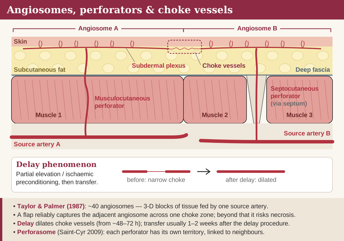
An angiosome is the three-dimensional block of tissue — skin, fat, fascia, muscle and bone — supplied by one source artery and its accompanying veins. Taylor and Palmer's injection studies mapped about 40 across the body, fitting together like a jigsaw in every layer, so the skin territory of a source artery overlies its deep territory.
Their anatomical observations still guide flap design. Vessels follow the connective tissue framework, travel with cutaneous nerves and radiate from fixed points such as septa and bony attachments; where one perforator is large its neighbours tend to be small, and vice versa. Venous territories mirror the arterial ones and are linked by avalvular ‘oscillating’ veins that allow flow in either direction across the boundary.
Choke vessels and true anastomoses
Adjacent angiosomes are connected in two ways: by choke vessels, which narrow at the boundary, and by true anastomoses, which cross between territories with no change in calibre. Choke vessels are the norm in skin, and their resistance is what limits how far a flap can be extended.
The working rule: a flap based on one angiosome reliably captures the adjacent angiosome across one choke zone, but the territory beyond a second choke zone is at risk of necrosis. Where territories are joined by true anastomoses, survival extends further. That is why partial flap loss is almost always distal, and why it reflects a design that outran its perfusion rather than a failed pedicle.
~40
angiosomes mapped by Taylor and Palmer
1
choke zone a flap reliably crosses
48–72 h
choke vessels begin to dilate after delay
10–14 d
approximate peak of the delay effect
Perforators: direct and indirect
Perforators reach the skin either directly, with skin supply as their main purpose, or indirectly, as spill-over branches of vessels whose main job is to feed muscle. Direct perforators — direct cutaneous and most septocutaneous vessels — tend to be larger, longer and quicker to dissect; indirect perforators are the musculocutaneous vessels that must be teased out through muscle. Terminology varies between authors, and the Gent consensus simply calls them septal and muscle perforators.
| Feature | Septocutaneous | Musculocutaneous |
|---|---|---|
| Course | Runs in an intermuscular septum, then pierces deep fascia | Passes through muscle before piercing deep fascia |
| Dissection | Usually quicker; pedicle lies in the septum | Intramuscular, slower and more variable |
| Typical flaps | Radial forearm, lateral arm, fibula skin paddle | DIEP, TDAP, most ALT perforators (through vastus lateralis) |
Perforasomes
A perforasome is the territory of a single perforator, and Saint-Cyr's work showed that each is linked to its neighbours by direct linking vessels in the fat and indirect linking vessels through the subdermal plexus. A flap raised on one perforator therefore captures neighbouring perforasomes through these links, filling those from the same source artery before those of an adjacent one.
- Orientate along the linking vessels. They run axially in the limbs and roughly perpendicular to the midline on the trunk, so skin paddles are designed in those directions.
- Flow direction is predictable. Perforators near a joint direct their flow away from it; those near the trunk midline flow away from the midline.
- Size the paddle to the perforator. A large perforator carries a large perforasome; a small one may need a second perforator to support a big paddle.
Mapping perforators before surgery turns these principles into a plan. A handheld Doppler is quick and cheap but cannot tell a perforator from an underlying axial vessel and over-calls signals. CT angiography shows calibre, the point where each vessel pierces the fascia and its intramuscular course, which is why it is widely used before abdominal perforator flaps; colour duplex ultrasound gives similar information without radiation but depends on the operator.
The delay phenomenon
Delay is a planned partial interruption of a flap's blood supply, made before transfer, that increases the area the flap can carry. Incising the margins or dividing a secondary pedicle forces flow through the remaining source, and the tissue adapts.
- Choke vessel dilatation. Choke vessels enlarge by hypertrophy and hyperplasia until they behave more like true anastomoses; this begins within about 48–72 hours.
- Reorientation of the vascular network along the flap's long axis.
- Sympathectomy effect. Dividing sympathetic fibres causes an early catecholamine surge followed by depletion and loss of vasoconstrictor tone.
- Ischaemic preconditioning. The tissue becomes metabolically more tolerant of ischaemia.
The effect is roughly maximal at 10–14 days, so transfer is usually planned about two weeks after the delay. Examples include the delayed pedicled TRAM, in which the deep inferior epigastric vessels are ligated in advance, and long random or distally based flaps.
Supercharging and turbocharging
Both techniques add a second inflow or outflow to a flap; the difference is where the extra vessel is joined. Supercharging anastomoses it to a recipient vessel at the defect — the deep inferior epigastric vessels of a pedicled TRAM joined to the thoracodorsal vessels, or the superficial inferior epigastric vein of a congested DIEP drained to a second recipient vein. Turbocharging joins it to the flap's own primary pedicle or a branch, so no extra recipient vessel is needed — classically a bipedicled abdominal flap in which the contralateral pedicle is anastomosed to a side branch of the ipsilateral one.
From territory to monitoring
Flap monitoring asks whether the pedicle is still flowing. Global compromise of the whole flap is a pedicle problem and needs urgent return to theatre; patchy distal loss in an otherwise healthy flap is a territory problem and is managed on its merits.
| Sign | Arterial insufficiency | Venous congestion |
|---|---|---|
| Colour | Pale | Blue or purple |
| Turgor | Flat, soft | Swollen, tense |
| Capillary refill | Slow or absent | Brisk |
| Pin-prick | Little or no bleeding | Rapid, dark bleeding |
Venous problems are more common than arterial ones, and most thromboses occur in the first 48–72 hours, which is when monitoring is most intensive. Clinical assessment is backed by handheld or implantable Doppler and, in some units, near-infrared spectroscopy; early re-exploration gives the best chance of salvage.
Frequently asked questions
What is a choke vessel?
A reduced-calibre vessel linking two adjacent angiosomes. It limits flow across the boundary, which is why a flap can usually capture only one neighbouring territory unless it is delayed or augmented.
What is the difference between an angiosome and a perforasome?
An angiosome is the whole three-dimensional territory of a source artery; a perforasome is the smaller territory of one perforator arising from it. Perforator flaps are designed on perforasomes.
How long after surgical delay should a flap be transferred?
Choke vessels begin to dilate within about 48–72 hours and the effect is roughly maximal at 10–14 days, so transfer is usually planned about two weeks after the delay.
What is the difference between supercharging and turbocharging?
Supercharging joins an extra flap vessel to a recipient vessel at the defect; turbocharging joins it to the flap's own pedicle or one of its branches. Both augment inflow or outflow.
Why is zone IV of an abdominal flap usually discarded?
It is the tissue furthest from the pedicle, beyond more than one choke zone, so its perfusion is the least reliable. Keeping it risks fat necrosis and partial flap loss.
References
- Janis JE (ed). Essentials of Plastic Surgery, 3rd ed. Thieme — chapters on flap physiology and microsurgery.
- Richards AM, Dafydd H. Key Notes on Plastic Surgery, 2nd ed. Wiley-Blackwell — Flaps; Microsurgery.
- Chung KC (ed). Grabb and Smith's Plastic Surgery, 8th ed. Wolters Kluwer — chapters on vascular territories and perforator flaps.
- Taylor GI, Palmer JH. The vascular territories (angiosomes) of the body: experimental study and clinical applications. Br J Plast Surg, 1987.
- Saint-Cyr M, Wong C, Schaverien M, Mojallal A, Rohrich RJ. The perforasome theory: vascular anatomy and clinical implications. Plast Reconstr Surg, 2009.
Further reading
- Angiosomes: Vascular Territories, Choke Vessels & Delay — thePlasticsFella — Independent open-access resource
- Delay Phenomenon — thePlasticsFella — Independent open-access resource
- Ischaemic & Congested Flap — thePlasticsFella — Independent open-access resource
Written for revision in the FRCS (Plast) style under our editorial standards. It is not clinical advice: always confirm against current guidance. Spotted an error? Tell us.
Related articles
General principles
Flap classification: blood supply, tissue and movement
Flap classification for FRCS (Plast): random vs axial, Mathes–Nahai types I–V, Cormack–Lamberty A–D, Nakajima, movement and Gent perforator nomenclature.
ReadGeneral principles
Local flap geometry: Z-plasty, rhomboid, bilobed and rotation flaps
Local flap design for FRCS (Plast): Z-plasty angles and gains, Limberg and Dufourmentel rhomboids, Zitelli bilobed, rotation, V–Y, keystone and forehead flaps.
ReadTurn reading into marks
Drill general principles with curriculum-mapped questions, rate your confidence, and watch the gap close on your dashboard.