General principles · Free article
Flap classification: blood supply, tissue and movement
Flap classification for FRCS (Plast): random vs axial, Mathes–Nahai types I–V, Cormack–Lamberty A–D, Nakajima, movement and Gent perforator nomenclature.
Three questions classify any flap
Every flap is classified by answering three questions: how is it perfused, what is it made of, and how does it reach the defect. A flap is tissue moved with a blood supply of its own — the property that separates it from a graft — which is why circulation dominates every scheme. Cormack and Lamberty's ‘five Cs’ (circulation, composition, contiguity, contour and conditioning) is a tidy checklist, but in the exam and in theatre the first three carry the weight.
Contiguity describes where the tissue comes from: local (adjacent to the defect), regional, distant pedicled (a groin flap to the hand, a cross-leg flap) or free, where the pedicle is divided and re-anastomosed at the recipient site.
Random versus axial supply
A random-pattern flap has no named vessel and survives on the dermal and subdermal plexus fed by unnamed perforators at its base; an axial-pattern flap carries a named artery and vein running along its long axis. McGregor and Morgan's distinction explains why a groin flap on the superficial circumflex iliac vessels can be raised far longer than a random flap of the same width.
The traditional length-to-breadth ratios for random flaps — about 1:1 on the trunk and limbs, more generous on the well-perfused face — are proxies for perfusion pressure, not laws. Milton's experiments showed that widening a random flap does not let you lengthen it: survival length is set by the pressure in the feeding vessels, and a wider base simply recruits more vessels at the same pressure.
Axial flaps can be peninsular, with a skin bridge left at the base, or islanded on the vessels alone, which frees them to rotate further. Classic examples are the paramedian forehead flap (supratrochlear vessels), the groin flap and the deltopectoral flap (internal mammary perforators).
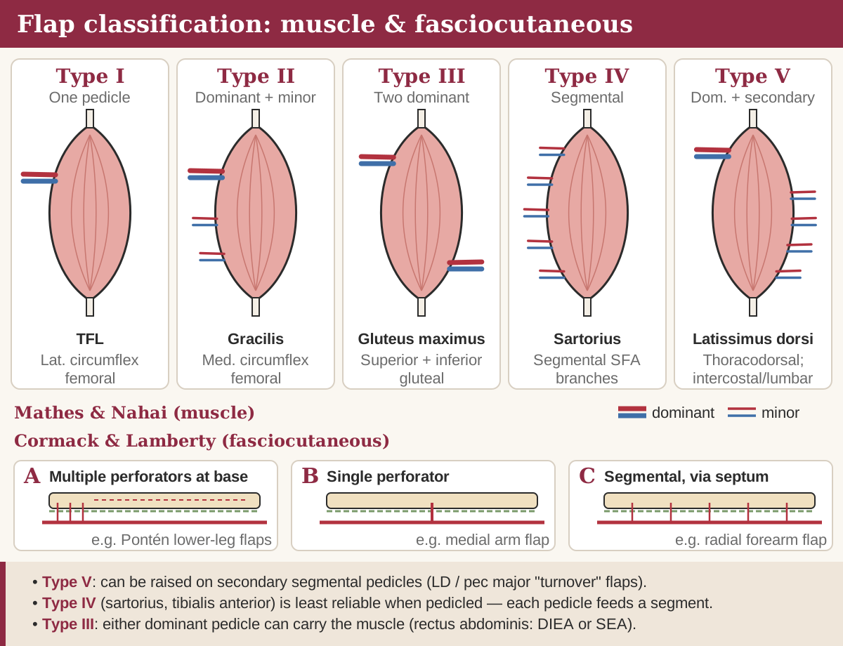
Muscle flaps: Mathes–Nahai types I–V
Mathes and Nahai classify muscles by the number and dominance of their vascular pedicles, and the type predicts how much muscle survives on any one pedicle. A dominant pedicle can carry the whole muscle; a minor pedicle keeps only part of it alive; segmental pedicles each supply their own slice.
| Type | Pattern | Classic examples | Clinical consequence |
|---|---|---|---|
| I | One vascular pedicle | Tensor fascia lata (lateral circumflex femoral); each gastrocnemius head (sural artery) | Reliable on its single pedicle; straightforward pedicled or free transfer |
| II | Dominant pedicle plus minor pedicles | Gracilis (medial circumflex femoral); soleus | Whole muscle survives on the dominant pedicle but not on a minor one; the most frequent pattern |
| III | Two dominant pedicles | Gluteus maximus (superior and inferior gluteal); rectus abdominis (superior and deep inferior epigastric); serratus anterior; the omentum (right and left gastroepiploic) | Survives on either pedicle and can be split into two flaps |
| IV | Segmental pedicles | Sartorius; tibialis anterior | Each pedicle supplies only its own segment, so the muscle is a poor flap if moved as a whole |
| V | One dominant plus secondary segmental pedicles | Latissimus dorsi (thoracodorsal; posterior intercostal and lumbar perforators); pectoralis major (thoracoacromial; internal mammary perforators) | Survives on either system, so it can be turned over on its segmental supply |
Fasciocutaneous flaps: Cormack–Lamberty A–D
Cormack and Lamberty grouped fasciocutaneous flaps by how their perforators reach the deep fascial plexus. Including the deep fascia captures the plexuses above and below it, which is why Pontén's lower-leg ‘superflaps’ survived at lengths that would kill a skin-only flap.
| Type | Vascular pattern | Example |
|---|---|---|
| A | Multiple fasciocutaneous perforators entering the base, orientated along the flap's long axis | Pontén lower-leg flaps |
| B | A single perforator of moderate size, so the flap can be islanded on it | Medial arm flap |
| C | Multiple small perforators reaching the fascia along an intermuscular septum from one source artery, which is harvested with the flap | Radial forearm flap |
| D | Type C extended to include adjacent muscle and bone (osteomyofasciocutaneous) | Radial forearm with a segment of radius; fibula with a skin paddle |
Nakajima extended the idea by describing six routes a deep fascial perforator can take from source vessel to skin — direct cutaneous, direct septocutaneous, direct cutaneous branch of a muscular vessel, perforating cutaneous branch of a muscular vessel, septocutaneous perforator and musculocutaneous perforator — and matching flap types to each. Its value is conceptual: it links the fasciocutaneous schemes to modern perforator flaps.
Composition and movement
Composition is what the flap carries: skin alone, fasciocutaneous, adipofascial, fascial, muscle, musculocutaneous, bone or osteocutaneous, or chimeric — several independent components, each on its own branch of a common pedicle. Add a sensory nerve and the flap is sensate; add a motor nerve to muscle and it becomes a functional transfer.
Movement describes how a local or pedicled flap reaches the defect; the geometry of each design is covered in Local flap geometry.
| Movement | What happens | Examples |
|---|---|---|
| Advancement | Slides forward in a straight line without rotation | V–Y, rectangular and bipedicle flaps; Moberg volar thumb flap |
| Rotation | Swings in an arc about a pivot point into an adjacent triangular defect | Scalp rotation; Mustardé cheek flap; gluteal rotation for sacral sores |
| Transposition | Pivots sideways into an adjacent defect; the donor site is closed separately | Rhomboid, bilobed, Z-plasty |
| Interpolation | Pivots over or under intact skin to a nearby defect; pedicle divided at a second stage | Paramedian forehead, nasolabial interpolation, cross-finger, Abbé |
| Distant pedicled or free | Carried from a remote site on a pedicle, or disconnected and re-anastomosed | Groin flap to the hand; free ALT, DIEP, fibula |
Perforator flaps and the Gent consensus
A perforator flap is skin and/or fat supplied by a perforator — a vessel that leaves its source artery and passes through or between deep tissues to reach the skin. The Gent consensus (Blondeel and colleagues, 2003) fixed the vocabulary: muscle perforators traverse muscle, septal perforators run only through an intermuscular septum.
- Name by vessel, not muscle. The flap takes the name of its nutrient artery — DIEAP (deep inferior epigastric artery perforator), TDAP (thoracodorsal), SGAP and IGAP (superior and inferior gluteal). Where more than one source vessel could supply a region, the region names the flap.
- Leave the muscle behind. Dissecting the perforator through the muscle spares the muscle and its motor nerve — the central argument for a DIEP over a TRAM.
- Free-style flaps are designed around any Doppler-located perforator rather than a pre-named template.
- The price is a longer, less forgiving dissection on variable anatomy, which is why CT angiography is widely used before a DIEP.
Delay, in brief
Surgical delay increases the area a flap can carry by dividing part of its supply and leaving the tissue in place before transfer. Choke vessels at the edge of the territory dilate and the tissue becomes more tolerant of ischaemia; the effect is near-maximal at about two weeks, which is when delayed transfers are usually scheduled. The mechanisms are covered in Angiosomes, perforasomes and delay.
Frequently asked questions
What is the difference between a random and an axial flap?
A random flap has no named vessel and survives on the subdermal plexus, so its safe length is limited. An axial flap carries a named artery and vein along its long axis and can be much longer for the same width.
Which Mathes–Nahai type is latissimus dorsi?
Type V. It has one dominant pedicle (thoracodorsal) and secondary segmental perforators from the posterior intercostal and lumbar vessels, so it survives on either system and can be used as a turnover flap.
Why is a type IV muscle a poor flap?
Its supply is segmental, so each pedicle nourishes only its own section. Moving the whole muscle on one pedicle risks losing the rest — sartorius and tibialis anterior behave this way.
How are perforator flaps named?
After the nutrient vessel, not the muscle the perforator passes through — for example DIEAP, TDAP or SGAP. If more than one source vessel could supply the area, the flap is named by region.
What type of flap is the radial forearm flap?
The classic Cormack–Lamberty type C fasciocutaneous flap: small septal perforators reach the fascia along the intermuscular septum, and the radial artery is harvested with the flap. Including radius makes it type D.
References
- Janis JE (ed). Essentials of Plastic Surgery, 3rd ed. Thieme — chapter on flap basics and classification.
- Richards AM, Dafydd H. Key Notes on Plastic Surgery, 2nd ed. Wiley-Blackwell — Flaps.
- Chung KC (ed). Grabb and Smith's Plastic Surgery, 8th ed. Wolters Kluwer — chapters on flap principles and perforator flaps.
- Mathes SJ, Nahai F. Classification of the vascular anatomy of muscles: experimental and clinical correlation. Plast Reconstr Surg, 1981.
- Cormack GC, Lamberty BGH. A classification of fascio-cutaneous flaps according to their patterns of vascularisation. Br J Plast Surg, 1984.
- Nakajima H, Fujino T, Adachi S. A new concept of vascular supply to the skin and classification of skin flaps according to their vascularization. Ann Plast Surg, 1986.
- Blondeel PN, et al. The ‘Gent’ consensus on perforator flap terminology: preliminary definitions. Plast Reconstr Surg, 2003.
Further reading
- Classification of Flaps — thePlasticsFella — Independent open-access resource
- Mathes and Nahai Classification — thePlasticsFella — Independent open-access resource
- Fasciocutaneous Flaps — thePlasticsFella — Independent open-access resource
Written for revision in the FRCS (Plast) style under our editorial standards. It is not clinical advice: always confirm against current guidance. Spotted an error? Tell us.
Related articles
General principles
Local flap geometry: Z-plasty, rhomboid, bilobed and rotation flaps
Local flap design for FRCS (Plast): Z-plasty angles and gains, Limberg and Dufourmentel rhomboids, Zitelli bilobed, rotation, V–Y, keystone and forehead flaps.
ReadGeneral principles
Angiosomes, perforasomes and the delay phenomenon
Taylor's angiosomes, choke vessels, Saint-Cyr's perforasomes, direct and indirect perforators, delay, supercharging and flap monitoring for FRCS (Plast).
ReadGeneral principles
Skin graft take: thickness, phases and failure
Skin grafts for FRCS (Plast): split vs full thickness, contraction, phases of take, recipient beds, causes of failure, meshing ratios, bolsters and NPWT.
ReadTurn reading into marks
Drill general principles with curriculum-mapped questions, rate your confidence, and watch the gap close on your dashboard.